Neil A. Switz
Education:
B.S., Physics, Stanford University
M.S., Applied & Engineering Physics, Cornell University
Ph.D., Biophysics, University of California Berkeley
Research
Optical imaging is central to enormous areas of science and technology, including mobile phone cameras to medical endoscopes to the Hubble Space Telescope. Optical microscopy has been central to biology for 350 years, and remains a primary method of medical diagnosis for many diseases, especially in remote and/or low-resource areas. My work tends to lie at the interface of optics and medical diagnostics, and benefits from my substantial industry experience; because I focus on deployable instruments that require actual
Typical student research projects involve fluorescence microscope design, photon and noise calculations for sensitive optical measurements, electronics and electronic design for photodetectors (including cameras), and developing approaches to computational optics and image processing.
My complete publication record can be found at ORCID; below are some examples of past and current projects:

COVID molecular diagnostic:
Fluorescence imaging combined with a molecular diagnostic technique known as CRISPR enabled rapid detection of COVID, and has the prospect of direct adaptation to many other diseases. Part of work with a large collaboration involving UC Berkeley and UCSF.
Recommended paper: “Amplification-free detection of SARS-CoV-2 with CRISPR-Cas13a and mobile phone microscopy,” Cell 2020. Publicity includes this, this, and this, among others.

Retinal imaging:
Technology we developed has also enabled handheld/portable fundus camera for retinal imaging and diagnosis of eye disease in emergency medicine environments as well as in low-resource settings – the latter key to helping reduce preventable causes of blindness.
Recommended paper:
“A Smartphone-Based Tool for Rapid, Portable, and Automated Wide-Field Retinal Imaging,” TVST 2018
Parasitic disease diagnosis:
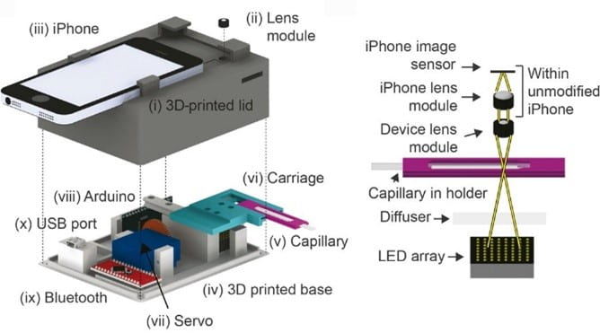
Onchocerciasis, known as “River blindness” is a major scourge in many countries, and the target of a worldwide eradication effort. The co-infection loa loa halted those efforts in Cameroon; our device – utilizing video microscopy and image analysis – has been used to diagnose > 50,000 people so far, and is an important component of restarting the eradication effort.
Recommended paper:
“Point-of-care quantification of blood-borne filarial parasites with a mobile phone microscope,” Science 2015. Publicity includes this, this, and this, among others.
Mobile-phone microscopy:
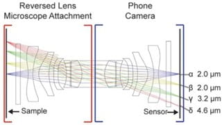
Mobile phone cameras, with their associated processing power, create the possibility of powerful, low-cost microscopes and diagnostic devices. I have been part of developing such devices for many years; our reversed-lens system enables ~ 2 micron resolution over a > 30 mm2 area, a huge improvement over previous systems.
Recommended paper:
“Low-Cost Mobile Phone Microscopy with a Reversed Mobile Phone Camera Lens,” PLOS One 2014.
Tuberculosis diagnosis:
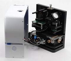
We designed a portable fluorescence microscope allowing extension of tuberculosis diagnosis outside of primary healthcare centers. These units were deployed for a year in Hanoi, Vietnam. We also developed an algorithm to automate diagnostic calls and reduce training requirements, a limiting factor in TB diagnosis.
Recommended paper:
“Mobile digital fluorescence microscopy for diagnosis of tuberculosis,” J. Clin. Microbiol. 2013.
My full publication record (see above for ORCID link) contains additional papers and patents – integral to commercial development of medical devices – on these and other (usually optical) projects.